CORE TEAM
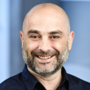
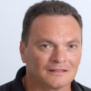
STÉPHANE COMPANT
Senior Scientist
AIT Austrian Institute of Technology
Dr. Stéphane Compant is Scientist working on plant-microbe interactions at the AIT. He received his PhD degree from the University of Reims Champagne-Ardenne and his habilitation from the University of Bordeaux in France. Stéphane Compant was Associate Professor of Microbiology at the National Polytechnic Institute of Toulouse in France before to be project leader at AIT. He is one of the leading research experts on microbial ecology of endophytic bacteria interacting with plants, on microscopy of plant-microbe interactions in general, and biocontrol of plant diseases using various biocontrol agents from different sources.
Microscopy of plant-microbe interactions.
Microorganisms living on and inside plants can be detected by various microscopy methods enabling to investigate their behaviour, their niches, and how they colonize their hosts. Different techniques are applied depending on whether single strains, full or synthetic microbiomes are studied. This talk will provide an introduction in the application of microscopy techniques to visualize adequately the microorganisms in their natural environment. Using these tools allows us to track down the microorganisms and to determine how they interact with the plants and how they are associated with various plant organs under different plant growth and environmental conditions.
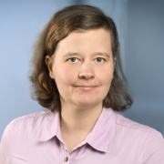
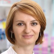
URSULA SAUER
Scientist
AIT Austrian Institute of Technology
Dr. Ursula Sauer received her Master in Biology at the University of Vienna and her PhD from the Vienna University of Technology. She is expert for the characterization of technical and biological surfaces and interfaces by means of fluorescence microscopy, scanning electron microscopy (SEM), and atomic force microscopy (AFM). A special focus of her work is on characterization of the phyllosphere and implications for plant ecology. Further, she is working on formulations for biocontrol agents as well as on development of environmentally sustainable formulations of plant growth promoting bacteria (PGPB).
AFM and SEM: close-ups of the phyllosphere
Plant cuticles are lipophilic membranes with protective function consisting of a polymer matrix covered with epicuticular waxes. Both, topography and chemistry control microhabitat quality in the phyllosphere and hence the abundance as well as the biodiversity of colonizing microbes. Atomic force microscopy (AFM) and Scanning Electron Microscopy (SEM) are excellent tools to study this habitat at the microscale.
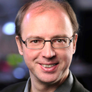
ADRIAN WALLNER
Scientist
AIT Austrian Institute of Technology
Dr. Adrian Wallner is a microbial ecologist. He has been focusing on bacterial communities with specific adaptions for the plant and fungal host environments. One of his main topics of interest are the bacterial strategies for endosymbiotic colonization and how they compare depending on the outcome of the interaction or the type of host that is colonized. Characterizing the levels of proximity between bacteria and hosts heavily relies on microscopy approaches which Dr. Wallner has applied on different models. Further characterization of the bacterial adaptations towards their host at the community level is achieved through amplicon sequencing and metagenomics approaches.
HANNA KOCH
Scientist
AIT Austrian Institute of Technology
Dr. Hanna Koch received her PhD degree at the University of Vienna focusing on metabolic versatility of nitrogen-cycling bacteria using different culture-dependent and independent techniques, including FISH and stable isotope labelling combined with single cell detection using NanoSIMS. During her PostDoc at the Radboud University, Hanna focused on different omics techniques to decipher the metabolic and phylogenetic diversity of different nitrifying groups. In addition, linking function and taxonomy became Hanna’s research passion. One method to answer the question “who is active in the environment under a specific condition” is BONCAT (BioOrthogonal Non-Canonical Amino acid Tagging), which labels translationally active microorganisms and which can combined with other downstream molecular techniques, such as FISH, or cell sorting followed by genome sequencing.
MARKUS GORFER
Scientist
AIT Austrian Institute of Technology
Dr. Markus Gorfer has put his research focus on functional ecology and the fungal contribution to environmental processes like heavy metal mobilization/immobilization, nutrient cycling and general ecosystem services. Modern techniques like qPCR, marker gene surveys, massive parallel sequencing and phylogenetic separation of environmental rRNA in conjunction with stable isotope probing are applied to gain insight into processes and players. As many environmentally important and dominant fungi can be cultivated, Dr. Gorfer has been developing molecular genetic tools to further research with hitherto often neglected organisms.
Transformation of fungi
Most fungi are amenable to genetic transformation, and the plant pathogenic bacterium Agrobacterium tumefaciens is widely used as donor for DNA-transfer into fungal cells. By this method, markers like fluorescent protein encoding genes can be introduced into host cells to allow microscopic visualization of tagged strains. Fungi expressing fluorescent proteins can be used for advanced colonization and interaction studies in an otherwise natural environment like plants that are normally colonized by a wide variety of prokaryotic and eukaryotic microorganisms. Coinocculations with several strains tagged with different colours are possible for advanced microbial interaction studies.
LEVI A. GHEBER
Scientist
Department of Biotechnology Engineering
Ben-Gurion University of the Negev
Dr. Gheber holds a Ph.D. in Physics of condensed matter (1995), followed by a 4 years postdoctoral training in biology in the Johns Hopkins University. He has joined the Department for Biotechnology Engineering at the Ben-Gurion University in 1999 and currently he serves as the Chairman of the Department. His lab conducts research on Nanobiotechnology and Biophysics. Dr. Gheber has published numerous articles in leading peer-reviewed journals, as well as several book chapters. He has served as an editorial board member for Biophysical Journal and is currently an editorial board member of Scientific Reports.
Atomic Force Microscopy for Biologists
The Atomic Force Microscope (AFM) was introduced in 1986 and has rapidly become an almost routine instrument in research labs. It owes its popularity to the fact that it can work on biological samples (unlike its “big brother”, the STM that was introduced 4 years earlier, and requires a conductive sample). The AFM enabled for the first time high resolution images on live biological specimens. We will discuss the components and modes of operation of AFM and will explain how the interactions between the AFM and the sample can create artifacts and how these can be avoided.
Image Analysis
The introduction of digital imaging has revolutionized the scientific world as much as it has, years later, revolutionized photography.
We will discuss properties of digital images and the files that hold them. We will get acquainted with digital image handling software and learn how to perform basic analysis and simple processing of digital images. These basic steps should allow the curious listener to launch on an autodidactic journey into more advanced topics.
MILICA PASTAR
Technician
AIT Austrian Institute of Technology
Mag. Milica Pastar holds a Master in Biology from the University of Banja Luka, Bosnia and Herzegovina, with a focus on Microbiology and Molecular Biology. She has long working experience in the microbiology laboratory of the Competence Centre for Bioresources at AIT. Her expertise ranges from daily routine microbiology methods to complex analysis of plant – microbe interactions, including fluorescence in situ hybridization (FISH).

